Long before the world had ever heard of covid-19, Kay Tye set out to answer a question that has taken on new resonance in the age of social distancing: When people feel lonely, do they crave social interactions in the same way a hungry person craves food? And could she and her colleagues detect and measure this “hunger” in the neural circuits of the brain?
“Loneliness is universal thing. If I were to ask people on the street, ‘Do you know what it means to be lonely?’ probably 99 or 100% of people would say yes,” explains Tye, a neuroscientist at the Salk Institute of Biological Sciences. “It seems reasonable to argue that it should be a concept in neuroscience. It’s just that nobody ever found a way to test it and localize it to specific cells. That’s what we are trying to do.”
In recent years, a vast scientific literature has emerged linking loneliness to depression, anxiety, alcoholism, and drug abuse. There is even a growing body of epidemiological work showing that loneliness makes you more likely to fall ill: it seems to prompt the chronic release of hormones that suppress healthy immune function. Biochemical changes from loneliness can accelerate the spread of cancer, hasten heart disease and Alzheimer’s, or simply drain the most vital among us of the will to go on. The ability to measure and detect it could help identify those at risk and pave the way for new kinds of interventions.
In the months ahead, many are warning, we’re likely to see the mental-health impacts of covid-19 play out on a global scale. Psychiatrists are already worried about rising rates of suicide and drug overdoses in the US, and social isolation, along with anxiety and chronic stress, is one likely cause. “The recognition of the impact of social isolation on the rest of mental health is going to hit everyone really soon,” Tye says. “I think the impact on mental health will be pretty intense and pretty immediate.”
Yet quantifying, or even defining, loneliness is a difficult challenge. So difficult, in fact, that neuroscientists have long avoided the topic.
Loneliness, Tye says, is inherently subjective. It’s possible to spend the day completely isolated, in quiet contemplation, and feel invigorated. Or to stew in alienated misery surrounded by a crowd, in the heart of a big city, or accompanied by close friends and family. Or, to take a more contemporary example, to participate in a Zoom call with loved ones in another city and feel deeply connected—or even more lonely than when the call began.
This fuzziness might explain the curious results that came back when Tye, before publishing her first scientific paper on the neuroscience of loneliness in 2016, ran a search for other papers on the topic. Though she found studies on loneliness in the psychological literature, the number of papers that also contained the words “cells,” “neurons,” or “brain” was precisely zero.
Neuroscientists have long assumed that questions about how loneliness might work in the human brain would elude their data-driven labs.
Though the nature of loneliness has preoccupied some of the greatest minds in philosophy, literature, and art for millennia, neuroscientists have long assumed that questions about how it might work in the human brain would elude their data-driven labs. How do you quantify the experience? And where would you even begin to look in the brain for the changes brought about by such a subjective feeling?
Tye hopes to change that by building an entirely new field: one aimed at analyzing and understanding how our sensory perceptions, previous experiences, genetic predispositions, and life situations combine with our environment to produce a concrete, measurable biological state called loneliness. And she wants to identify what that seemingly ineffable experience looks like when it is activated in the brain.
If Tye succeeds, it could lead to new tools for identifying and monitoring those at risk from illnesses worsened by loneliness. It could also yield better ways to handle what could be a looming public health crisis triggered by covid-19.
Finding the loneliness neurons
Tye has homed in on specific populations of neurons in rodent brains that seem to be associated with a measurable need for social interaction—a hunger that can be manipulated by directly stimulating the neurons themselves. To pinpoint these neurons, Tye relied on a technique she developed while working as a postdoc in the Stanford University lab of Karl Deisseroth.
Deisseroth had pioneered optogenetics, a technique in which genetically engineered, light-sensitive proteins are implanted into brain cells; researchers can then turn individual neurons on or off simply by shining lights on them though fiber-optic cables. Though the technique is far too invasive to use in people—as well as an injection into the brain to deliver the proteins, it requires threading the fiber-optic cable through the skull and directly into the brain—it allows researchers to tweak neurons in live, freely moving rodents and then observe their behavior.
Tye began using optogenetics in rodents to trace the neural circuits involved in emotion, motivation, and social behaviors. She found that by activating a neuron and then identifying the other parts of the brain that responded to the signal the neuron gave out, she could trace the discrete circuits of cells that work together to perform specific functions. Tye meticulously traced the connections out of the amygdala, an almond-shaped set of neurons thought to be the seat of fear and anxiety both in rodents and in humans.

Scientists had long known that stimulating the amygdala as a whole could cause an animal to cower in fear. But by following the maze of connections in and out of different parts of the amygdala, Tye was able to demonstrate that the brain’s “fear circuit” was capable of imbuing sensory stimuli with far more nuance than previously understood. It seemed, in fact, to modulate courage too.
By the time Tye set up her lab at MIT’s Picower Institute for Learning and Memory in 2012, she was following the neural connections of the amygdala to places like the prefrontal cortex, known as the brain’s executive, and the hippocampus, the seat of episodic memory. The goal was to construct maps of the circuits across the brain that we rely on to understand the world, make meaning of our moment-to-moment experience, and respond to different situations.
She began studying loneliness largely by serendipity. While scouting for new postdocs, Tye came across the work of Gillian Matthews. As a graduate student at Imperial College London, Matthews had made an unexpected discovery when she separated the mice in her experiments from one another. Social isolation—the very fact of being alone—seemed to have changed brain cells called DRN neurons in ways that implied they might play a role in loneliness.
Tye immediately saw the possibilities. “Oh, my gosh—this is incredible!” she recalls thinking. That the signs of social isolation could be traced to a specific part of the brain made total sense to her. “But where is it and how would you find it? If this could be the region, I thought, that would be super interesting.” In all her studies of neurons, says Tye, “I’d never seen anything about social isolation before. Ever.”
Tye realized that if she and Matthews could construct a map of a loneliness circuit, they could answer in the lab precisely the kinds of questions she hoped to explore: How does the brain imbue social isolation with meaning? How and when does the objective experience of not being around people, in other words, become the subjective experience of loneliness? The first step was to better understand the role the DRN neurons played in this mental state.
DRN neurons are shown here within the dopamine system and downstream circuitry.
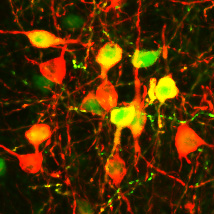
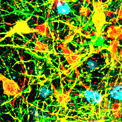
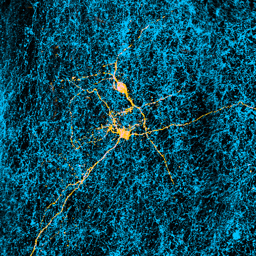
One of the first things Tye and Matthews noticed was that when they stimulated these neurons, the animals were more likely to seek social interaction with other mice. In a later experiment, they showed that animals, when given the choice, actively avoided areas of their cages that, when entered, triggered the activation of the neurons. This suggested that their quest for social interaction was driven more by a desire to avoid pain than to generate pleasure—an experience that mimicked the “aversive” experience of loneliness.
In a follow-up experiment, the researchers put some of the mice in solitary confinement for 24 hours and then reintroduced them to social groups. As one would expect, the animals sought out and spent an unusual amount of time interacting with other animals, as if they’d been “lonely.” Then Tye and Matthews isolated the same mice again, this time using optogenetics to silence the DRN neurons after the period in solitary. This time, the animals lost the desire for social contact. It was as if the social isolation had not been registered in their brains.
Scientists have long known that the brain harbors the biological equivalent of a car’s fuel gauge—a complex homeostatic system that allows our gray matter to track the state of our basic biological needs, like those for food, water, and sleep. The purpose of the system is to drive us toward behaviors aimed at maintaining or restoring our natural state of balance.
Tye and Matthews seemed to have found the equivalent of a homeostatic regulator for the basic social-contact needs of rodents. The next question: What do these findings mean for people?
Hungry for a smile
To answer that question, Tye is working with researchers in the lab of Rebecca Saxe, a professor of cognitive neuroscience at MIT, who specializes in the study of human social cognition and emotion.
The human experiments are far harder to design because the brain surgery required for optogenetics is not an option. But it’s possible to expose lonely people to pictures of friendly people offering social cues—like a smile—and then monitor and record changes in blood flow to different parts of the brain using fMRI imaging. And, thanks to previous experiments, scientists have a good idea of where in the brain to look—an area analogous to the one Matthews and Tye studied in mice.
Last year, Livia Tomova, a postdoc who has been overseeing the research in Saxe’s lab, recruited 40 volunteers who self-identified as having large social networks and very low levels of loneliness. Tomova exiled her subjects to a room in the lab and forbade any human contact for 10 hours. For comparison, Tomova asked the same participants to come back for a second 10-hour session that contained plenty of social interaction, but no food.
Tomova and Saxe used fMRI scans to measure the brain’s response to food and social interaction after periods of fasting and isolation. The scan on the right shows activity in the midbrain associated with rewards.
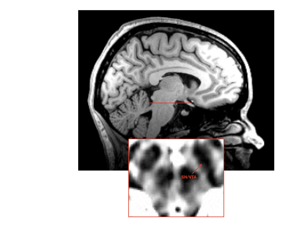
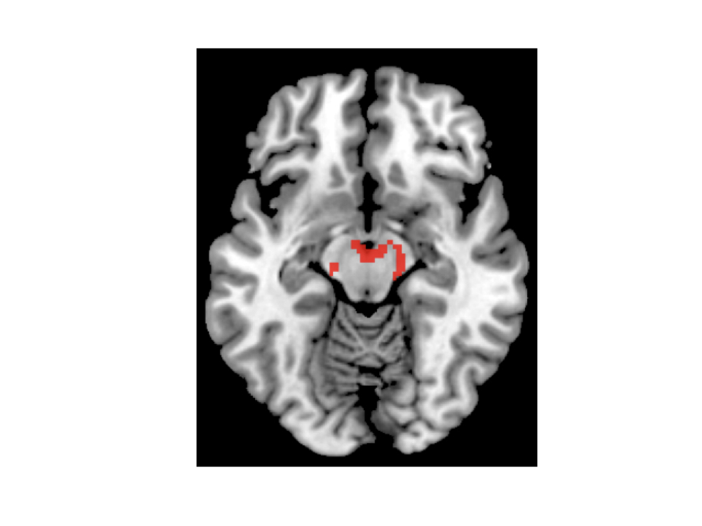
At the end of each session, the subjects were asked to climb into the fMRI scanner and were exposed to different images: some showed people offering nonverbal social cues, and others contained pictures of food.
Unlike Tye and Matthews, Tomova was unable to home in on individual neurons. But she was able to track changes in blood flow within larger areas of the scan, known as voxels; each voxel displayed the changing activity of discrete populations of several thousand neurons. Tomova focused in on areas of the midbrain known to be rich in the neurons associated with producing and processing the neurotransmitter dopamine.
These areas have already been linked in other experiments to the sensation of “wanting” or “craving” something. They’re areas that light up in response to images of food when a person is hungry, or to drug-related images in people with addiction. Would they do the same in lonely people shown pictures of a smile?
The answer was clear: after the social isolation, the subjects’ brain scans showed far more activity in the midbrain when they were shown the pictures of social cues. When the subjects were hungry but had not been socially isolated, they exhibited a similarly robust reaction to the food cues, but not to the social ones.
“Whether it’s the drive for social contact or the drive for other things like food, it seems to be represented in a very similar way,” says Tomova.
The pandemic experiment
Understanding how the hunger for social contact is produced in the brain might allow a deeper understanding of the role social isolation plays in some illnesses.
Objectively measuring loneliness in the brain, as opposed to asking people how they feel, could provide some clarity on the connection between depression and loneliness, for example. Which comes first—does depression cause loneliness, or does loneliness cause depression? And might social intervention applied at the right time help fight depression?
Insights into the circuitry of loneliness in the brain might also shed some light on addiction, which isolated animals are more prone to, according to some research. The evidence appears particularly strong in adolescent animals, which seem to be even more sensitive to the effects of social isolation than older or younger ones. Humans between the ages of 16 and 24 are the most likely to report feeling lonely, and this is also the age when many mental-health disorders first begin to manifest. Is there a connection?
Insights into the circuitry of loneliness in the brain might shed some light on addiction.
But the most obvious current need may be in response to the social isolation prompted by the covid pandemic. Some internet surveys report no overall increase in loneliness since the pandemic began, but what about people at most risk of mental-health problems? When they are isolated, at what point does it begin to endanger their psychological and physical well-being? And what types of interventions might protect them from that danger? Once we can measure loneliness, we can begin to find out, making it far easier to design targeted interventions.
“A vital question for future research is how much, and what kinds of positive social interaction are sufficient to fulfill this basic need and thus eliminate the neural craving response,” Tomova and Tye wrote in a preprint of their forthcoming paper, posted at the end of March. The pandemic “emphasized the need for a better understanding of human social needs and the neural mechanism underlying social motivation,” they wrote. “The current study provides a first step in that direction.”
That, in the typically understated language of science, signals the birth of a whole new field of research—not something you often get to witness, let alone be part of.
“It’s just so exciting to me, because these are all concepts that we’ve heard about a million times in psychology, and for the very first time, we actually have cells in the brain that we can link to the system,” Tye says. “And once you have one cell, you can trace backwards, you can trace forwards; you can figure out what’s upstream; you can figure out what all the neurons that are upstream are doing, and what messengers are being sent,” says Tye. “Now you can find the whole circuit; you know where to start.”
from MIT Technology Review https://ift.tt/2QVtlfO

No comments:
Post a Comment